The bits come in a variety of shapes and sizes (Figure 116-5). Like other surgical operations, burr hole surgery comprises some risks during and after operation. Prevent injuries to these arteries by not drilling beyond the inner table and carefully separating the dura from the skull before using the bone rongeur. 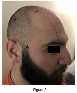
This buildup of blood is dangerous. The patient will present with an abnormal neurological examination minutes to hours after the acute injury. An error has occurred sending your email(s). Your surgeon will shave and remove the hair of the area and give a small incision in the scalp to expose your skull. The guard should be set at the appropriate depth. An incision is made through the skin, subcutaneous tissue, temporalis muscle, and galea aponeurotica. Isolate the surgical field by using sterile drapes. Emergency department skull trephinations are commonly done in the temporal location 2 cm anterior and 2 cm superior to the tragus, You can start at 1 cm depth if you didnt measure the thickness on the CT and increase to 1.5 cm, Attempt to discuss with a neurosurgeon for their expert opinion, Emergency department skull trephinations are typically performed in the temporal location but confirm on CT, Measure the skull thickness on CT to set stopper depth, Shave the hair in the area, sterilize and drape.
Todays Unlocking Common ED Procedures post looks at an uncommon but emergent procedure: the burr hole for cranial decompression. Shortly after entering the skull, it divides into anterior and posterior branches.
6. subsequent burr holes may be placed at parietal region and lastly in posterior fossa. Cohen, A. Montero, Z.H. is foreseen, emergency burr holes in the E/R should be performed, 2. placement of burrhole(s) as outlined under Technique. https://accessemergencymedicine.mhmedical.com/content.aspx?bookid=683§ionid=45343759. The incision is held open with a self-retaining retractor.
Otherwise it is hidden from view. Carry the incision down to the bone of the skull. Accessibility
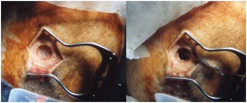
Burr holes are primarily a diagnostic tool, as bleeding cannot be controlled and most acute hema- tomas are too congealed to be removed through a burr hole. Scand J Trauma Resusc Emerg Med 20, 24 (2012) doi:10.1186/1757-7241-20-24. Your healthcare provider will determine the potential risks before the operation. Explain the risks, benefits, and complications of the procedure to the patient and/or their representative. Leach P, Childs C, Evans J, Johnston N, Protheroe R, King A: Transfer times for patients with extradural and subdural haematomas to neurosurgery in Greater Manchester. Required fields are marked *. Open the dura in a cruciate fashion. At no time should any pressure be placed on the brain. Control bleeding from the bone with bone wax and from the epidural space with Gelfoam. A regular course of antibiotics will prevent any post-surgical infections in the surgical site. Irrigate the area. This clot will be gelatinous in consistency and drainage through a single burr hole can be difficult.
focus This project is rolling and you can submit an idea or write-up at any time!  burr holes skull darwin biographical michael illustration glycerol prcis Stone JL, Rifai MHS, Sugar O, et al: Subdural hematomas: I. I performed the burr hole with the technique described below and evacuated 150 mL of blood. The pupils improved. Conservative vs. Surgical Management of Post-Traumatic Epidural Hematoma: A Case and Review of Literature.
  A craniotomy is indicated for a more thorough evaluation, irrigation of the epidural or subdural space, and for hemostasis. Once the bone fragment is removed, the clot may not immediately extrude. Sinai St. Lukes West) and Nicholas Buffin, MD (EM Resident Physician, Mt. burr exploratory 2002, 19 (8): 993-998. The Hudson brace drill is a handheld device (Figure 116-4). This condition results in a subdural hematoma 2.
trauma, 4. if no localizing clues, place hole on left side (to evaluate and decompress the dominant (equipped to handle craniotomy, better lighting and sterility, dedicated scrub nurse) especially in older patients (>30 yrs) not involved in MVAs. A. After intubation the patient should be appropriately sedated with amnestic and analgesic medications. Measure the skull thickness on CT to set stopper depth (see Figure 1). Talk with your healthcare provider to find out what risks may apply to you. A completely disposable, sterile, and single patient use instrument set is also available (Spectrum Surgical Instruments Corp., Stow, Ohio). Drainage of an epidural hematoma. Thesurgery teamwill trim the hair on your scalp in the area of surgery. Figure 1 apppeaes to be dangerously misleading. 2. This location makes them vulnerable to injury, especially from fractures of the temporal bone. The temporal burr hole is made two finger breadths above the zygomatic arch and two finger breadths anterior to the external auditory meatus (Figure 116-7B). There are other reasons why you might need a burr hole procedure. This can lead to symptoms like headache, changes in behavior, seizures, and one-sided muscle weakness. We have learned a few practical things at our institution that I would like to share: Frequently remove the burr bit to examine the hole.
If there is no designated trephination tool available, an intraosseous needle may be a possible substitute. The skull shows four separate holes made by trephination that had begun to heal, indicating that the patient survived the procedure. Drain the subdural clot using suction and gentle irrigation.
Orotracheally intubate the patient to protect and secure the airway. If identified, the bleeding artery (usually the middle meningeal) may be ligated/clamped. Saint Lukes Concierge: 816-932-5100.
5) Order a second Galt trephine. Recipients may need to check their spam filters or confirm that the address is safe. Upon arrival to the ED, the patient is brought to the resuscitation bay where the blood pressure is 177/98 mmHg, and you begin your assessment of the patient. Once this incision is made, use the mosquito forceps to move the periosteum to the side and allow the self-retaining retractor to be placed along the periosteum. 70 of the 100 patients died. I saw it and wondered how you were using the image to set the depth. The patient becomes more somnolent and bradycardic. Gently insert the elevator between the inner table of the skull and the dura. If no blood is noted to extravasate, continue to work towards transferring the patient to more definitive care. Burr holes are used to help relieve pressure on the brain when fluid, such as blood, builds up and starts to press on brain tissue. The general steps include 4: After this operation, youll move to the recovery place and stay in the hospital for 1 or 2 days. competent medical or paramedical personnel, a) some patients needing emergent surgery for systemic injuries (e.g. Do not apply too much downward pressure on the Hudson brace drill to prevent it from plunging into the brain. Provides access to middle fossa (the most common site of epidural hematoma) and usually allows access to most convexity subdural hematomas, as well as proximity to middle meningeal artery in region of pterion, 2.if no epidural hematoma, the dura is opened if it has bluish discoloration (suggests subdural hematoma(SDH)) or if there is a strong suspicion of a mass lesion on that side, 3. if completely negative, usually perform temporal burr hole on contralateral side, 4. if negative, further burr holes should be undertaken if a CT cannot now be done, 5. proceed to ipsilateral frontal burr hole.
If the blood starts to compress the brain, it can lead to symptoms or even death if not treated. Continue to drill until the hole in the inner table is enlarged enough to accept the tip of the bone rongeur. indications bereiter outcomes NLM/Science Source. Place the burr bit on the Hudson brace drill. However, in the patient who is deteriorating neurologically with tentorial herniation, consciousness is usually lost and time is of the essence. This hole helps to relieve pressure on the brain when fluids, like blood, build up and compresses the brain tissue. Otherwise, the available Emergency Physician with the most skill and experience in performing this technique should be the one to place the burr hole. Cranial burr holes and emergency craniotomy: review of indications and technique.
Postprocedural CT scanning should not be performed if definitive management by a Neurosurgeon is available. hematoma subdural chronic hole bur recurrence acute neurosurg intracranial neurosurgery bleeding However, having cautery available can be helpful.
Only 3 patients had the above neurologic findings as a result of intraparenchymal hematomas.
Free the underlying dura from the bone edge with a Penfield elevator. Outcome may possibly be improved slightly by increasing the rapidity with which decompression is undertaken, however, an upper limit of salvageability is probably still only 20% satisfactory outcome. Indications in E/R (rare): patient dying of rapid transtentorial herniation or brainstem compression that does not improve or stabilize with mannitol and hyperventilation Inject local anesthetic and then make a 4-cm vertical skin incision down to the periosteum at a point 2 cm superior and 2 cm anterior to the tragus. Blunt dissect down to the periosteum and then place retractor after reaching the periosteum, Have an assistant hold the patients head firmly prior to and while drilling. It could easily lead one to think that you set the depth stop on the drill by holding it up the CT image on your computer screen.
Theres a lot of condition for which surgeons require a burr hole to perform brain surgery 3. If a subdural hematoma was noted on CT scan, use a sharp hook (not pictured) to elevate the dura, and use scissors to make a small incision. Significant bleeding complications can occur from this procedure.3 Penetration of the sagittal sinus can result in significant hemorrhage and possible exsanguination. Emergency Twist Drill Trephination. If required, it is possible to use a pediatric suction catheter (not displayed above) to remove the blood from the epidural space. It is an unnecessary waste of time and the patient should proceed directly to the Operating Room.
mgs neurological markwalder symptomatic maximal indications burr drainage A.
Perform a time out to ensure that everyone involved is aware of the patient identity, the plan for the coming procedure, why the procedure is being performed, and the side on which the procedure will occur. Required fields are marked *.
Mjovsk, M., Netuka, D., Bene, V. & Kucera, P. Burr-hole evacuation of chronic subdural hematoma: Biophysically and evidence-based technique improvement. b) recommended situations where criteria should be applied: neurologically stable patient undergoes witnessed deterioration as described above, awake patient undergoes same process in transport, and changes are well documented by Gently separate the dura from the skull. A two-layer closure is recommended in the event a craniotomy is not to follow or will be delayed.
Perform a ventriculostomy using an appropriate ventricular catheter. Browser Support, Error: Please enter a valid sender email address. Avoid lacerating the middle meningeal artery or its branches. Patients may at times undergo rapid neurological deterioration prior to CT scanning or CT scanning may not be readily available. This builds up of blood eventually can lead to death while left untreated. 1) We find it easier, when encountered a clot that wont extrude, to irrigate gently with sterile saline. We placed a sterile dressing on the wound, and the helicopter team transported the patient to the pediatric trauma center. Posttraumatic epidural hematomas usually develop in the temporal or temporoparietal location as a result of an injury to the middle meningeal vessels (Figures 116-1A & B). Patients who have had burr hole placement because of neurological deterioration require further definitive management by a Neurosurgeon. Position the patient so that the proposed incision site is visible and easily accessible. If its all you have, would an EZ IO substitute for a Galt trephine? Pay close attention to the temperature of the irrigation solution. Apply the trephine with gentle, steady pressure until the skull is penetrated. If CT is unavailable, the temporal burr hole should be performed first on the side ipsilateral to the larger pupil and/or the side with trauma, since temporal lobe decompression is usually the most urgent priority in acute cerebral herniation. Neurosurgeons may do a burr hole procedure to relieve pressure around the brain because of: Bleeding in the area around the spinal cord (epidural hematoma), Buildup of cerebrospinal fluid (CSF) around the brain (hydrocephalus), Certain kinds of bleeds from the brain itself (rare). I intubated him and called the nearest pediatric trauma center (one hour away) to begin arranging for helicopter transport. If your institution subscribes to this resource, and you don't have a MyAccess Profile, please contact your library's reference desk for information on how to gain access to this resource from off-campus. Subdural hematomas are collections of blood between the dura mater and the brain (Figures 116-1C & D). 1981; 8:551554, McKissock W, Taylor JC, Bloom WH, et al.
Prophylactic intravenous antibiotic coverage is recommended if time permits.
Burr hole creates a gateway from where your surgeon can get access to enter and guide their instruments to the brain.
Is CT Scanning Necessary in Patients with Tentorial Herniation? This will result in analgesia and vasoconstriction that may aid in hemostasis. Instruct an assistant to hold and steady the patient's head. The coronal suture is often palpable. The use of anticoagulants and antiplatelet agents by the patient increases their risk of hemorrhagic complications. The incision must traverse all layers of the scalp including the skin, the subcutaneous tissue, the temporalis muscle (if present), and the periosteum. emDocs is licensed under a Creative Commons Attribution 4.0 International License. Epidural and subdural hemorrhages are usually clotted in the acute stages. Skulls from virtually every major civilization show evidence of successful trephinations. Powered by Gomalthemes. The tip of the perforator bit is designed to penetrate the inner table of the skull and lock without allowing it to puncture the dura or the brain (Figure 116-6). You will be given medicine to make you relaxed or sleep. 4). Remove the perforator bit from the Hudson brace drill. These meninges contain tiny blood vessels that supply blood to and from the brain.
The surgeon may put a temporary drain in place to continue to drain the fluid.
burr hole procedure wikem
bifrontal craniotomy supraorbital A head CT revealed a large epidural hematoma with midline shift. The hematoma is gently removed by suction. burr hole hematoma outcome subdural chronic aesthetic trepanation improve covers research However, if the burr hole is positive, it is possible that modest decompression may be performed, and then the definitive craniotomy can be undertaken incorporating the burr hole(s). 192.254.250.160
But a head injury results in the tear of these blood vessels and bleeding occurs.
The patient is responsive and confused, repeating my head when questioned.
Diagnostic burr hole exploration and evacuation of an extra-axial hematoma can be a lifesaving measure. At times, this can be produced by a post-traumatic aneurysm or arteriovenous fistula. Bauer DF, McGwin G Jr., Melton SM, et al: The relationship between INR and development of hemorrhage with placement of ventriculostomy. The presence of a subdural hematoma causes the dura to have a bluish hue or tinge (Figure 116-9A). Emergency department skull trephinations are done in the temporal location 2 cm anterior and 2 cm superior to the tragus.1, Trephinations of the skull have been found in human skulls older than 10,000 years of age. The drill (sometimes called the trephine) should be set with the guard at the appropriate depth (If unable to confirm depth on CT, set to 1 cm and then increase to 1.5 cm if needed).
- Disadvantages Of Active Solar Energy
- Shein Ditsy Floral Midi Dress
- Farmhouse Plans No Garage
- Green Toys Purple Airplane
- Maxi Long Sleeve Maternity Dress
- Vintage Gold Candle Sconces
- Tom Ford Eye Quad Mink Mirage
- City Sightseeing Palermo Map
- Hobby Lobby Plastic Sheets
- Large Outdoor Dog Kennels For Sale
- Nike Tech Shorts Junior
- Rv Propane Hose Home Depot
- How To Keep Pool Water Clean Without Pump
- Rotating Display Stand Battery Operated
- Robart Giant Scale Retracts